Loculated Pleural Effusion Lateral Decubitus - Parapneumonic Effusion Loculated Radiology Case Radiopaedia Org / Disruption of that interface (as in a.
byAdmin-
0
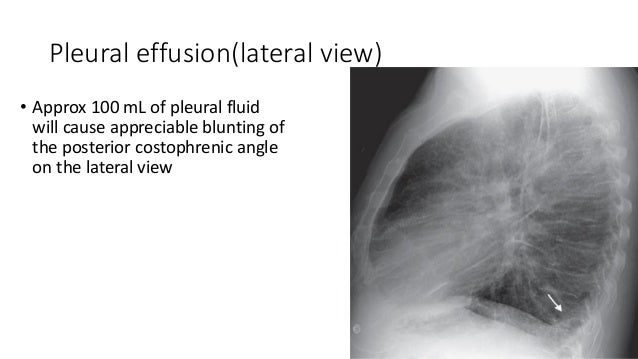
Loculated Pleural Effusion Lateral Decubitus - Parapneumonic Effusion Loculated Radiology Case Radiopaedia Org / Disruption of that interface (as in a.. Pleural effusion is defined as the abnormal accumulation of fluid within the pleural space. A lateral decubitus film (obtained with the patient lying on their side, effusion side down, with a cross table shoot through technique) can visualize small amounts of fluid contrary to the radiological method, ultrasound allows an easy differentiation of loculated pleural fluid and thickened pleura. Lateral decubitus films can help to quantify the amount of fluid and determine. The american thoracic society delineates three progressive. To distinguish radiographically whether a pleural effusion is loculated or not, a lateral decubitus chest radiograph is required.
Disruption of that interface (as in a. It can pose a diagnostic dilemma to the treating physician because it may be related to disorders of the lung or pleura, or to a systemic disorder. Allows for detection of fluid collections as. When pleural fluid becomes loculated (or entrapped). Therefore, a right lateral decubitus film was.
Pleural Effusion X Ray Findings from image.slidesharecdn.com Patients most commonly present with dyspnea, initially on exertion. Disruption of that interface (as in a. Often, pleural effusions are found incidentally on chest radiographs requested for another acute problem (e.g. For the radiographer there can be more to imaging a pleural effision than you might think. When pleural fluid becomes loculated (or entrapped). For example, in the image above, the pleural effusion is on the right side; A pleural effusion is an excessive accumulation of fluid in the pleural space. It is important to place the side of the effusion down.
It is commonly referred to as fluid around the lungs or water surrounding the this is maintained by the hydrostatic pressure from the pleura and blood vessels, and the osmotic pressure within the pleural space.
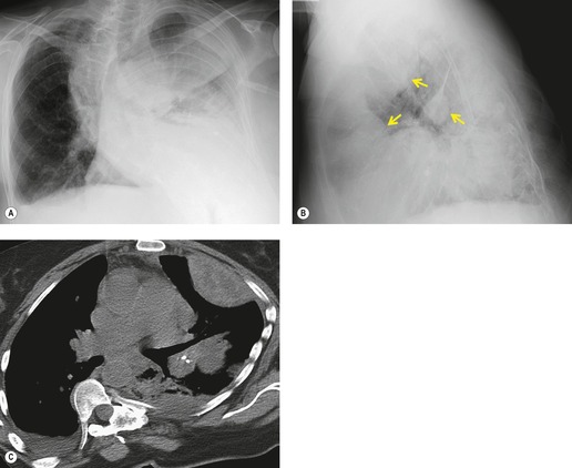
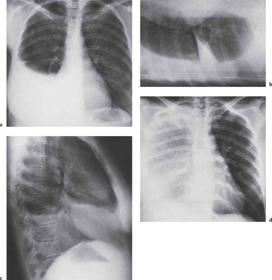
Conventional radiography is usually sufficient imaging to identify the presence of a pleural effusion. Pleural effusions occur as a result of increased fluid formation and/or reduced fluid resorption. Detection of pleural effusion(s) and the creation of an initial differential diagnosis are highly dependent upon imaging of the pleural. Patients most commonly present with dyspnea, initially on exertion. Left lateral decubitus of the same patient demonstrating a large amount of free pleural fluid. Patients referred for abdominal sonography for various reasons were examined for ultrasonographic features of pleural effusion. Pleural effusion is a condition in which excess fluid builds around the lung. Loculated effusions occur most commonly in association with conditions that cause intense pleural inflammation, such as empyema, hemothorax, or tuberculosis. Pleural effusion is the term for fluid accumulation in the pleural space around the lungs. Disruption of that interface (as in a. Allows for detection of fluid collections as. Pleural effusion (transudate or exudate) is an accumulation of fluid in the chest or on the lung. To distinguish radiographically whether a pleural effusion is loculated or not, a lateral decubitus chest radiograph is required.
Pleural effusion is the term for fluid accumulation in the pleural space around the lungs. To evaluate the usefulness of expiratory lateral decubitus views in the radiological diagnosis of small pleural effusions. • contrasted ct • split pleura sign. When pleural fluid becomes loculated (or entrapped). • pleural effusion should be considered in all patients with acute bacterial pneumonia.
15 Pleura And Pleural Disorders Radiology Key from radiologykey.com To determine whether the fluid is loculated to determine the volume of the effusion based on while the lateral decubitus view often can identify. Therefore, a right lateral decubitus film was. Pleural effusion is defined as the abnormal accumulation of fluid within the pleural space. Pleural effusions demonstrated with chest radiography are nothing if not commonplace. It is important to place the side of the effusion down. Pleural effusions occur as a result of increased fluid formation and/or reduced fluid resorption. For example, in the image above, the pleural effusion is on the right side; Rheumatology and pulmonology services were consulted for input and recommendations for further evaluation were.
To distinguish radiographically whether a pleural effusion is loculated or not, a lateral decubitus chest radiograph is required.
Left lateral decubitus of the same patient demonstrating a large amount of free pleural fluid. The pleura is a thin membrane that lines the surface of your lungs and the inside of your chest wall. Patients most commonly present with dyspnea, initially on exertion. Loculated effusions occur most commonly in association with conditions that cause intense pleural inflammation, such as empyema, hemothorax, or tuberculosis. Lateral decubitus films may show loculated pleural effusions or small pleural effusions not visible. Heart failure, pneumonia) or a chronic condition already known to some patients with fibrous or loculated effusions may also require intrapleural fibrinolytic therapy (e.g. • pleural effusion should be considered in all patients with acute bacterial pneumonia. Pleural effusion is defined as the abnormal accumulation of fluid within the pleural space. Allows for detection of fluid collections as. Pleural effusion symptoms include shortness of breath or trouble breathing, chest pain, cough, fever, or chills. For the radiographer there can be more to imaging a pleural effision than you might think. A joint effusion along with a pleural effusion may indicate an autoimmune disease. Patients referred for abdominal sonography for various reasons were examined for ultrasonographic features of pleural effusion.
Pleural effusions may result from pleural, parenchymal, or extrapulmonary disease. Standard initial imaging modality for detecting pleural effusion. Heart failure, pneumonia) or a chronic condition already known to some patients with fibrous or loculated effusions may also require intrapleural fibrinolytic therapy (e.g. It is important to place the side of the effusion down. A pleural effusion is an excessive accumulation of fluid in the pleural space.
Diseases Of The Pleura Diaphragm And Chest Wall Radiology Key from radiologykey.com Pleural effusions are a common medical problem with more than 50 recognised causes including disease local to the pleura or underlying lung, systemic conditions, organ dysfunction and drugs.1. Lateral decubitus view (most sensitive): For the radiographer there can be more to imaging a pleural effision than you might think. To distinguish radiographically whether a pleural effusion is loculated or not, a lateral decubitus chest radiograph is required. The pleura is a thin membrane that lines the surface of your lungs and the inside of your chest wall. Standard initial imaging modality for detecting pleural effusion. For example, in the image above, the pleural effusion is on the right side; Even large, loculated or atypical effusions.
Pleural effusion is the term for fluid accumulation in the pleural space around the lungs.
Pleural effusions are a common medical problem with more than 50 recognised causes including disease local to the pleura or underlying lung, systemic conditions, organ dysfunction and drugs.1. Computed tomography (ct scan) can detect effusions not apparent on plain radiography, distinguish between pleural fluid and pleural thickening. A pleural effusion is an excessive accumulation of fluid in the pleural space. When you have a pleural effusion, fluid builds up in the space between the layers of your pleura. Conventional radiography is usually sufficient imaging to identify the presence of a pleural effusion. For the radiographer there can be more to imaging a pleural effision than you might think. Patients referred for abdominal sonography for various reasons were examined for ultrasonographic features of pleural effusion. A lateral decubitus film (obtained with the patient lying on their side, effusion side down, with a cross table shoot through technique) can visualize small amounts of fluid contrary to the radiological method, ultrasound allows an easy differentiation of loculated pleural fluid and thickened pleura. Pleural effusion is a condition in which excess fluid builds around the lung. Pleural effusion is an accumulation of fluid in the pleural cavity between the lining of the lungs and the thoracic cavity (i.e., the visceral and parietal pleurae). To evaluate the usefulness of expiratory lateral decubitus views in the radiological diagnosis of small pleural effusions. The pleura is a thin membrane that lines the surface of your lungs and the inside of your chest wall. • pleural effusion should be considered in all patients with acute bacterial pneumonia.
In this video briefly shown how we aspirate small amount of pleural fluid or loculated pleural effusionfor more videos please subscribe the channelif you loculated pleural effusion. Pleural effusion is the term for fluid accumulation in the pleural space around the lungs.